Medicine
Deep dive – cancer diagnostics
PUBLISHED IN Deep Dive Articles: APRIL 2024
Cancer diagnostic and the current landscape
Cancer diagnostics involve the identification and confirmation of the presence, type, and characteristics of cancer in an individual. Early and accurate diagnosis is crucial for effective treatment and improved outcomes. Various methods and techniques are employed in cancer diagnostics, and the choice of diagnostic approach depends on factors such as the type of cancer suspected, the patient’s medical history, and the symptoms observed.
Medical history and physical examination
Physicians often start with a thorough review of the patient’s medical history and conduct a physical examination to identify any signs or symptoms of cancer.
Imaging techniques
X-rays: Basic X-ray imaging can be used to identify abnormalities in certain organs or bones.
Computed Tomography (CT) Scan: Provides detailed cross-sectional images of the body, allowing for the visualization of tumours and their size.
Magnetic Resonance Imaging (MRI): Uses powerful magnets and radio waves to create detailed images, particularly useful for soft tissues like the brain and muscles.
Ultrasound: Uses sound waves to create images and is commonly used for breast, abdominal, and pelvic examinations.
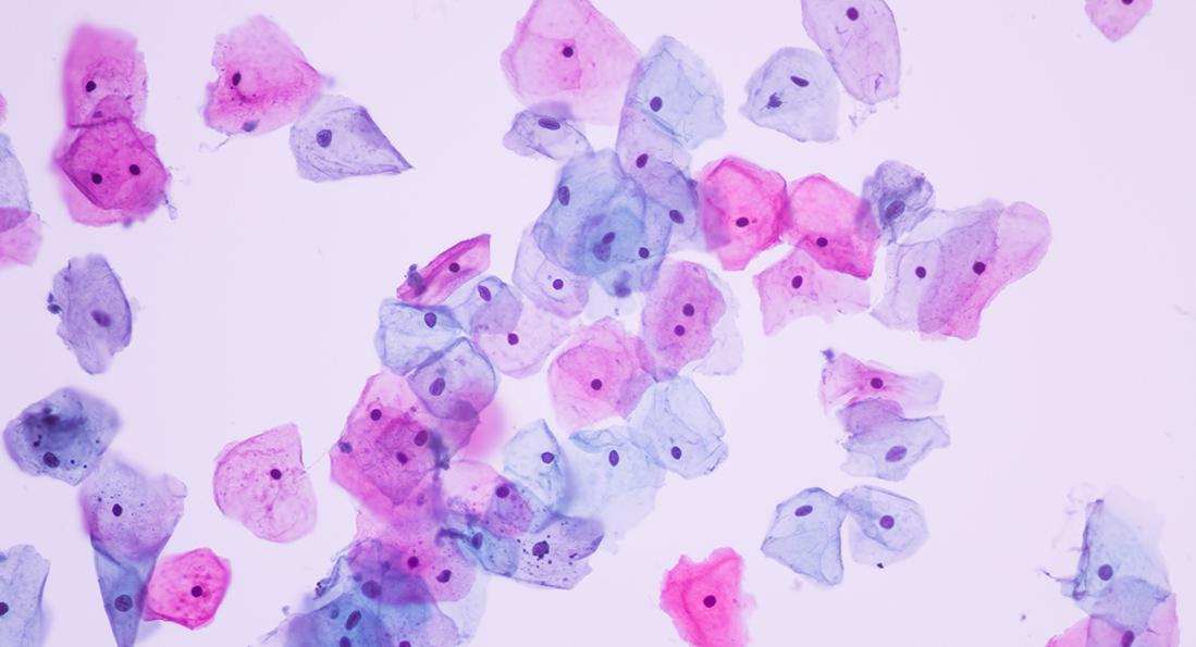
Biopsy
The definitive method for cancer diagnosis involves the removal of a small tissue sample (biopsy) from the suspected tumour site. This tissue is then examined under a microscope by a pathologist to determine if cancer is present, and if so, to identify its type and characteristics.
Blood tests and tumour markers
Blood tests may be conducted to look for specific substances (tumour markers) that are produced by cancer cells. Elevated levels of these markers can indicate the presence of cancer, although they are not conclusive on their own and may require further investigation.
Endoscopy
A procedure in which a flexible tube with a camera on the end is used to visually inspect the inside of organs like the digestive tract, lungs, or bladder. Biopsies can also be taken during endoscopy.
Genetic testing
Analysing a person’s DNA to identify specific genetic mutations or alterations that may increase the risk of certain cancers or indicate the presence of cancer.
Positron emission tomography (PET) scan
A nuclear medicine imaging technique that helps identify areas of increased metabolic activity, which can be indicative of cancer.
Liquid biopsy
A relatively newer technique that involves analysing blood samples for circulating tumour cells or fragments of tumour DNA to detect and monitor cancer.
New frontiers for cancer diagnostics
Advancements in technology and research have led to several new frontiers in cancer diagnostics, offering improved accuracy, earlier detection, and personalized treatment approaches. Here are some notable developments in cancer diagnostics:
Liquid biopsies
Liquid biopsies involve analysing blood or other body fluids for traces of cancer-related genetic material, such as circulating tumour DNA (ctDNA) or circulating tumour cells (CTCs). This non-invasive method allows for real-time monitoring of cancer, early detection of recurrence, and assessment of treatment response.
Artificial Intelligence (AI) and Machine Learning
AI and machine learning algorithms are being increasingly utilized in the interpretation of medical imaging, such as radiology and pathology slides. These technologies can help identify subtle patterns and anomalies that may not be easily discernible by human observers, improving the accuracy of cancer detection and classification.
Genomic profiling
Comprehensive genomic profiling examines the entire DNA of a tumour to identify specific genetic alterations. This information helps guide treatment decisions, as targeted therapies can be matched to the unique genetic profile of the cancer.
Multiparametric imaging
Combining different imaging modalities, such as MRI, CT, and PET scans, enables a more comprehensive evaluation of tumours. This approach provides a more detailed and accurate assessment of tumour characteristics, aiding in treatment planning and monitoring.
Liquid biopsy for early detection
Researchers are exploring the potential of liquid biopsies for the early detection of cancer, even before symptoms manifest. Identifying genetic markers or other molecular changes associated with early-stage cancers can enable prompt intervention and improved outcomes.
Metabolomics
Metabolomics involves the study of the unique metabolic signatures associated with cancer. By analysing the small molecules present in tissues or biofluids, researchers can gain insights into the metabolic changes associated with different types of cancer, potentially leading to new diagnostic markers.
Circulating tumour cells (CTCs) analysis
The isolation and analysis of CTCs from blood samples provide information about the characteristics of cancer cells, allowing for a better understanding of the disease’s progression and the potential for personalized treatment strategies.
Molecular imaging
Molecular imaging techniques, such as positron emission tomography (PET) with specific tracers, enable the visualization of molecular and cellular processes within the body. This helps in detecting and characterizing tumours at a molecular level.
Exosome analysis
Exosomes are small vesicles released by cells, including cancer cells, into the bloodstream. Analysing the contents of exosomes can provide information about the tumour microenvironment, treatment response, and the presence of specific biomarkers.
Smartphone-based diagnostics
Research is ongoing to develop low-cost, portable devices and smartphone applications for cancer diagnostics. These technologies aim to bring diagnostic capabilities to resource-limited settings and facilitate early detection.